Services provided by the Translational Pathology Imaging Laboratory include:
- Panel design and optimization (6 antibodies + DAPI)
- Automated multiplex staining on the Leica BOND RX
- Slide Imaging on the PhenoImager HT
- Region of interest selection
- Image analysis with inForm® – includes tissue/cell segmentation algorithms and quantification of individual biomarkers and phenotypes
- Additional custom analyses – biomarker co-expression, spatial relationships between cellular phenotypes
- inForm® workstation availability for independent use
Access the TPIL Intake Form. Email Maron Joy, PhD (joym@upmc.edu), with completed intake form to schedule initial project meeting.
Multiplex Staining and Imaging Workflow
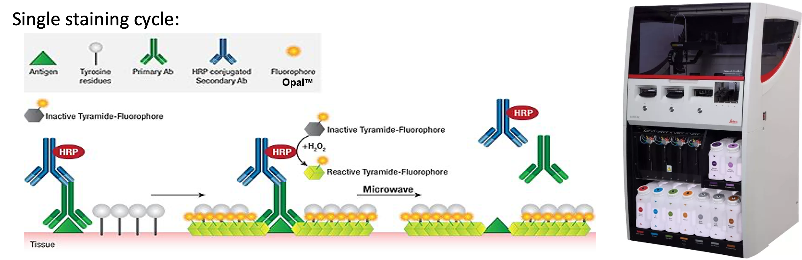
1. Sequential staining of seven markers on the BOND RX.
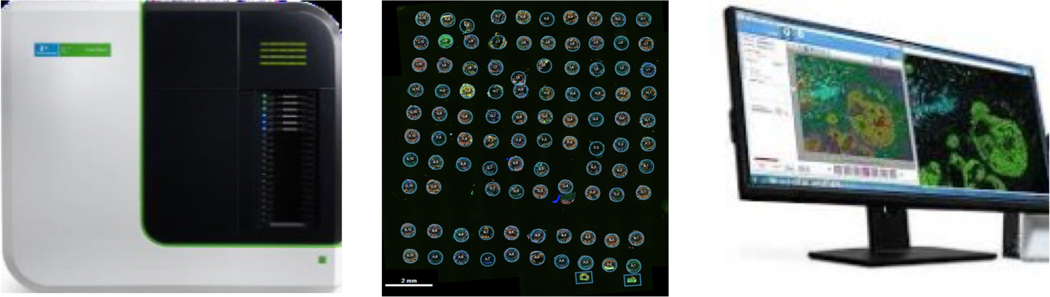
2. Whole slide scanning with the PhenoImager HT
3. Select regions of interest in Phenochart
4. Analysis of ROIs using inForm/phenoptrReports

